2007年9月30日 星期日

Shock Index


References:
  • Rady MY, Smithline HA, et al. A comparison of the shock index and conventional vital signs to identify acute, critical illness in the Emergency Department. Ann Emerg Med. 1994; 24: 685-690.
  • Yealy DM, Delbridge TR. The shock index: All that glitters. Ann Emerg Med. 1994; 24: 714-715.

Mallampati test

The patient sits in front of the anaesthetist and opens the mouth wide.
The patient is assigned a grade according to the best view obtained.


View obtained during Mallampati test:
1. Faucial pillars, soft palate and uvula visualised
2. Faucial pillars and soft palate visualised, but uvula masked by the base of the tongue
3. Only soft palate visualised
4. Soft palate not seen.

Clinically, Grade 1 usually predicts an easy intubation and Grade 3 or 4 suggests a significant chance that the patient will prove difficult to intubate.

The results from this test are influenced by the ability to open the mouth, the size and mobility of the tongue and other intra-oral structures and movement at the craniocervical junction.

Cormack and Lehane classification


Class I: the vocal cords are visible
Class II the vocals cords are only partly visible
Class III only the epiglottis is seen
Class IV the epiglottis cannot be seen.

2007年9月26日 星期三

Purple Urine


2007年9月24日 星期一

CPAS

  • A simple method for obtaining temporary pelvic stabilization when an external fixator cannot be applied is the application of a circumferential pelvic anti-shock sheet (CPAS).
  • Advantages of this technique include the fact that it is inexpensive and readily available in all EDs. Special training is not required and the emergency physician can apply the sheet. Lower extremity and abdominal access is maintained after the sheet is placed.
  • Caution is required in patients with lateral compression pelvic ring injuries or sacral neuroforaminal fractures. Forceful or aggressive CPAS application could worsen visceral injury or sacral nerve root injury in these instances.


Circumferential pelvic anti-shock sheeting. A. A sheet is placed under the pelvis. B. The ends are brought together anteriorly. C. Hemostats are used to secure the sheet snugly.

2007年9月23日 星期日

Estimated Blood Volume

Estimated Blood Volume 之算法:
  • PREMATURE = 100 cc/kg
  • INFANT = 90 cc/kg
  • CHILD = 80 cc/kg
  • MEN = 70 cc/kg
  • WOMEN = 60 cc/kg

何時要做 Urine Alkalinization?

Indication: Drug Overdose with weak acid
  • Phenobarbital or barbiturate overdose
  • All Class I antidysrhythmic overdose
  • Other sodium channel blockers
  • Tricyclic Antidepressant (TCA) Overdose
  • Antihistamine Overdose
  • Cocaine Overdose
  • Salicylates
  • Chlorpropamide (Diabenese)
  • Sulfonamides (some)
  • Methotrexate
  • Fluoride
  • Diflunisal

2007年9月21日 星期五

2007年9月20日 星期四

看 ICH:CT 還是比 MRI 略勝一籌

Magnetic Resonance Imaging and Computed Tomography in Emergency Assessment of Patients with Suspected Acute Stroke: A Prospective Comparison.
 
Chalela JA, et al. Lancet 2007;369:293

BACKGROUND: MRI is believed to be more sensitive than CT scanning for imaging ischemic stroke, but is considered insufficiently sensitive for acute hemorrhagic stroke.

METHODS: In this single-center study at a suburban hospital in Bethesda, MD, and coordinated at the National Institutes of Health, 356 patients with suspected acute strokes underwent MRI and CT scanning of the brain. The imaging studies were later reviewed by a panel of two neuroradiologists and two stroke neurologists blinded to the patients' clinical findings.

RESULTS: The final diagnosis was acute ischemic stroke in 190 patients (53%), acute intracranial hemorrhage in 27 (8%) and transient ischemic attack in 14 percent. Twenty-five percent of the patients were not ultimately diagnosed with cerebrovascular disease. The sensitivity of CT scanning was 26% for acute stroke overall, 16% for acute ischemic stroke and 89% for acute intracranial hemorrhage (corresponding specificities 98%, 98% and 100%, respectively). The sensitivity of MRI was 83% for acute stroke overall, 83% for acute ischemic stroke and 81% for acute intracranial hemorrhage (corresponding specificities, 97%, 96% and 100%, respectively). In the subgroup of patients imaged within three hours of symptom onset, sensitivities for acute stroke were 27% for CT scanning and 76% for MRI, and specificities were 100% and 96%, respectively.

CONCLUSIONS: Although the authors suggest that their findings are consistent with a superiority of MRI over CT scanning for the imaging of acute stroke, MRI missed more patients with acute hemorrhagic stroke, which could have significant implications when selecting patients for thrombolytic therapy.

--
張志華 敬上

2007年9月19日 星期三

Emphysematous Cholecystitis


Yogesh Shrestha, M.D.
Steven Trottier, M.D.
Saint Louis University

2007年9月18日 星期二

小兒 appendicitis 會診前可以給 morphine 止痛

最新的 RCT:會診前給 morphine 不影響 小兒 appendicitis 手術之決定
Journal Home
Efficacy and Impact of Intravenous Morphine Before Surgical Consultation in Children With Right Lower Quadrant Pain Suggestive of Appendicitis - A Randomized Controlled Trial

Received 15 April 2007; accepted 19 April 2007. published online 28 June 2007.

Study objective
The evidence supporting the use of analgesia in children with abdominal pain suggestive of appendicitis is limited. The objectives of the study are to evaluate the efficacy of morphine before surgical consultation in children presenting to the pediatric emergency department (ED) with right lower quadrant pain suggestive of appendicitis and determine whether it has an impact on the time between arrival in the ED and the surgical decision.

Methods
All children between the ages of 8 and 18 years who presented to a pediatric ED with a presumptive diagnosis of appendicitis were eligible to be enrolled in a randomized double-blind placebo-controlled trial if the initial pain was at least 5 of 10 on a verbal numeric scale. Patients received either 0.1 mg/kg of intravenous morphine (maximum 5 mg) or placebo. The primary outcomes were (1) the difference in pain using a visual analog scale at baseline and 30 minutes after the completion of the intervention, analyzed by comparing the mean pain differences for the treatment versus placebo groups; and (2) the time between arrival in the ED and the surgical decision, analyzed by comparing the median delay for the 2 groups.

Results
Ninety patients with a suspected diagnosis of appendicitis were randomized to receive morphine or placebo. Both groups were similar in terms of demographics, medical history, physical findings, emergency physician assessment of the probability of appendicitis, and initial pain score. There was no important difference in the decrease of pain between the morphine (n=45) and placebo (n=42) groups 30 minutes after the intervention: 24±23 mm and 20±18 mm, respectively (Δ 4 mm [95% confidence interval [CI] −5 to 12 mm]). There was also no important difference in the time between arrival in the ED and the surgical decision: median 269 minutes (95% CI 240 to 355 minutes) for morphine and 307 minutes (95% CI 239 to 415 minutes) for placebo (Δ −34 minutes [95% CI −105 to 40 minutes]).

Conclusion
The use of morphine in children with a presumptive diagnosis of appendicitis did not delay the surgical decision. In our group of patients, however, morphine at a dose of 0.1 mg/kg was not more effective than placebo in diminishing their pain at 30 minutes.

2007年9月17日 星期一

Idraparinux for DVT

治癒 DVT 的長效藥物:Idraparinux


ILMA 小技巧


這個小技巧我以前不知道,今天讀到跟大家分享大家知道ILMA會配備專用的ETT,好處是夠硬(有內建鋼圈)但價錢當然也非同小可,因此很多時候都是使用一般的ETT來放今天介紹的trick是當你要放入普通ETT的時候,記得把curve倒過來(專用ETT沒有curve,是直的)這樣子穿出來的時候的角度就是B圖上面的樣子,比較容易放入trachea裡面

----
侯勝文

2007年9月14日 星期五

Low-Tidal-Volume Ventilation

Low-tidal-volume ventilation should be implemented at an initial tidal volume of 6 ml per kilogram of predicted, not actual, body weight, as in the ARDSNet trial (NEJM, 2000). The predicted body weight (PBW) is calculated as follows:


  • for men, PBW = 50.0 + 0.91 (height in centimeters - 152.4); and

  • for women, PBW = 45.5 + 0.91 (height in centimeters - 152.4).

Lung size has been shown to depend most strongly on height and sex. A given person should not receive a higher lung volume just because of weight gain as the lungs are essentially the same size irrespective of weight gain.
Figure 2. Conventional Ventilation as Compared with Protective Ventilation.
NEJM: This example of ventilation of a 70-kg patient with ARDS shows that conventional ventilation at a tidal volume of 12 ml per kilogram of body weight and an end-expiratory pressure of 0 cm of water (Panel A) can lead to alveolar overdistention (at peak inflation) and collapse (at the end of exhalation). Protective ventilation at a tidal volume of 6 ml per kilogram (Panel B) limits overinflation and end-expiratory collapse by providing a low tidal volume and an adequate positive end-expiratory pressure. Adapted from Tobin.

Permissive hypercapnia

Low-tidal-volume ventilation can result in an increase in the partial pressure of carbon dioxide to above the normal range (permissive hypercapnia).
What are the risks and benefits of permissive hypercapnia?
Answer:
Permissive hypercapnia can result in respiratory acidosis, which can be mitigated to some extent by increasing the respiratory rate and gradual renal buffering of the acidosis. Potentially harmful consequences of permissive hypercapnia include pulmonary vasoconstriction and pulmonary hypertension, proarrhythmic effects of increased discharge of the sympathetic nervous system, and cerebral vasodilation, yielding increased intracranial pressure.
However, experimental data have suggested that permissive hypercapnia is not only safe but also potentially beneficial. In most cases, hemodynamic characteristics actually improve, owing to the release of catecholamines.
Nonetheless, permissive hypercapnia should probably be used with caution in patients with heart disease and is relatively contraindicated in patients with elevated intracranial pressure.

Acute Lung Injury

Acute lung injury (ALI) is defined as the acute onset of impaired gas exchange (the ratio of the partial pressure of arterial oxygen in millimeters of mercury to the FiO2 of <300) and the presence of bilateral alveolar or interstitial infiltrates in the absence of congestive heart failure.

Common causes of ARDS are
  1. sepsis (with or without a pulmonary source),
  2. trauma, aspiration,
  3. multiple blood transfusions,
  4. pancreatitis,
  5. inhalation injury, and
  6. certain types of drug toxicity.

2007年9月13日 星期四

Herpetic glossitis


別誤以為是 candidiasis...
From: NEJM

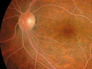
眼底病變

Test #1.


請問答案是??
請反白:『Lipemia retinalis』。
---------------------------------------------------------------
Test #2
請問答案是??
請反白:『CRAO with Cherry red spot』。

2007年9月12日 星期三

Sudden Hearing Loss

What is Sudden Hearing Loss?
Sudden hearing loss (SHL) is defined as greater than 30 dB hearing reduction, over at least three contiguous frequencies, occurring over a period of 72 hours or less. Some patients describe that the hearing loss was noticed instantaneously in the morning and others report that it rapidly developed over a period of hours or days. The severity of the hearing loss however varies from one patient to another and only one ear is usually affected. There have been some reports of involvement of both ears with SHL. Tinnitus is usually reported in patients with SHL loss and vertigo can be present in 40% of cases. The incidence of SHL has been reported to be 5-20 per 100,000 person per year and accounts for 1% of all sensorineural hearing loss cases. Males are equally affected as females. The average age at onset is reported to be 46 to 49 years with increasing incidence with age.

What Causes Sudden Hearing Loss?
There are many causes for sudden hearing loss which include infectious, circulatory, inner ear problems like meniere’s disease, neoplastic, traumatic, metabolic, neurologic, immunologic, toxic, cochlear, idiopathic (unknown cause) and other causes. Unfortunately, even after a thorough search for a possible pathology, the cause of sudden hearing loss remains unknown in most patients.

How is Sudden Hearing Loss Diagnosed?
Evaluation usually begins with a careful history and physical examintion looking for potential infectious causes such as otitis media, systemic diseases and exposure to known ototoxic medications. In essence, SHL is diagnosed by documenting a recent decline in hearing. This generally requires an audiogram. Blood studies are usually performed in an attempt to rule potentially systemic causes of SHL including syphilis, Lyme disease, metabolic, autoimmune, and circulatory disorders. Magnetic resonance imaging (MRI) of the brain is recommended to rule out an acoustic neuroma which is reported to be existent up to 15% of patients with sudden hearing loss.

How is Sudden Hearing Loss Treated?
Due to the lack of a definite cause of sudden hearing loss, its treatment has been controversial. Over the years, this has included systemic steroids, antiviral medications, vasodilators, carbogen therapy either (alone or in combination) or no treatment at all. The no treatment option was based on the high reported rate of spontaneous recovery up to two third of cases.

2007年9月5日 星期三

醫療法

請各位要熟知醫療法:
  • 第60條:不得拒絕處理危急病患;
  • 第81條:要善盡病情告知義務。

-------------------------------------

-------------------------------------